DIAGNOSIS AND X-RAY IMAGING
Every individual’s oral health varies, and as a result, the dentist will evaluate your needs and recommend an X-ray schedule accordingly. If you’re a new patient, the dentist may advise taking a full series of X-rays or panoramic image to assess your current oral health state, and use this as a baseline going forward.
Correct diagnosis can be made by correct xray imaging.There are two main types of dental X-rays:
Intraoral X-rays
Intraoral X-rays are the most common type of dental X-ray taken. These X-rays provide a lot of detail and allow your dentist to find cavities, check the health of the tooth root and bone surrounding the tooth, check the status of developing teeth, and monitor the general health of your teeth and jawbone.
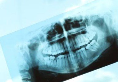
Extraoral X-rays
Extraoral X-rays show teeth, but their main focus is the jaw and skull. These X-rays do not provide the detail found with intraoral X-rays and therefore are not used for detecting cavities or for identifying problems with individual teeth. Instead, extraoral X-rays are used to look for impacted teeth, monitor growth and development of the jaws in relation to the teeth, and to identify potential problems between teeth and jaws and the temporomandibular joint or other bones of the face.
Follow Us!